Room 3: Diagnosis
 The diagnosis of epilepsy in Greek-Byzantine medicine
 Epilepsy diagnosis in ancient Rome
 modern Methods for diagnosing epilepsy
The diagnosis of epilepsy in Greek-Byzantine medicine
The Greek physician Alexandros of Tralleis (525-605) suggested the following method to diagnose epilepsy: 'Wash the head of the patient and burn a ram's horn under his nose and he will fall down.' (In ancient times the goat was considered to be the mammal which was most prone to epileptic seizures.)
Epilepsy diagnosis in ancient Rome
In the Roman era it was usual to give people suspected of having epilepsy a piece of jet to smell. If the person did not fall to the ground on smelling the stone, he was considered to be 'free of the falling sickness'. (This was for a time a common procedure when buying slaves.)
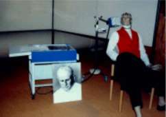
A similar test was undertaken using a potter's wheel. It was believed that a person with epilepsy would fall to the ground on watching the wheel turn. It is possible that people who actually did fall to the ground during this test actually did so as a result of their photosensibility. Flashing lights or glittering surfaces can trigger epileptic seizures in some people. (Today seizures can be provoked by computer games, the television or the lights in a disco.)
Modern methods for diagnosing epilepsy
Today, electroencephalography (a method of measuring brain wave patterns, often abbreviated to EEG) plays a decisive role in diagnosing epilepsy.
 The human electroencephalogram was developed by the German psychiatrist
Hans Berger (1873-1941) in the 1920s.
Every nerve cell (neuron) is a highly complex form whose function is linked to electrical (and chemical) processes. With the help of an EEG, the electrical processese can be made visible in a very simplified form. A piece of EEG equipment is basically an instrument which receives and intensifies electrical potentials (voltage fluctuations) and transfers them to a recording instrument (which is now in many cases a computer).
The fluctuations of the electrical impulses can be measured outside the skull
using pairs of electrodes (e.g. of silver
chloride). Each pair of electrodes transmits a signal to the EEG, consisting of the
difference of the voltage between the pair and showing the cumulative potential of thousands
of neurons. These fluctuations vary within a range of less than a millionth of a volt.
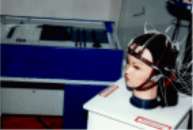
 The electrodes are placed on the head of the patient following an internationally laid-down
pattern and held in place with a rubber cap, or stuck to the scalp. Depending on the
condition and head-size of the patient, 20, 40, or more electrodes can be used.
Each electrode is connected to its pair (bipolar) or to an inert reference electrode (unipolar).
A normal EEG varies depending on the age of the patient and whether he or she is awake or asleep.
The EEG of a patient with a cerebral disorder often looks different. In patients with epilepsy, the EEG often (but not always) has characteristic differences. However, if a patient has a normal EEG, it does not prove that that person does not have epilepsy.
Computer-Tomography (CT: a method for measuring differences in density in organic tissue using X-rays) is another technique used to diagnose epilepsy as is magnet-resonance-imaging (MRI: a diagnosis technique using strong magnetic fields).

